Release time :2023-11-02
Source:support@yingchitech.com
Scan:1590
Clinical Support Department of Shenzhen Yingchi Technology Co.,Ltd.
Children and adolescents, a stage of significant brain development and hormonal fluctuations, exhibit different TMS responses. This complexity extends into adulthood, where aging-related neurophysiological changes, such as cortical atrophy and changes in cortical excitability, further alter TMS responses. In the previous article, we summarized the impact of neurodevelopment on TMS detection indicators and rTMS treatment. This article continues to summarize the impact of neurodegenerative processes on TMS detection indicators and rTMS treatment.


As individuals enter physiological aging, complex biological changes occur at the molecular, cellular, and systemic levels.
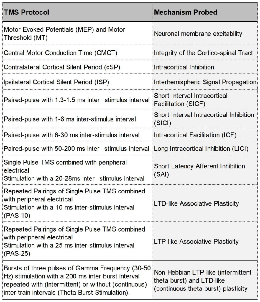
①RMT: Age-related cortical atrophy can increase the distance from the coil to the cortex, making RMT a valid measure for examining brain aging. Several studies and meta-analyses have reported increased RMT in older adults, but there are also inconsistent results.
②SICI: Studies have been inconsistent. A meta-analysis comparing 187 older adults with 169 younger adults showed reduced SICI in older adults, but the difference was not statistically significant.
③ICF: This is normal in most studies, with a few exceptions. A meta-analysis showed a slight but non-significant decrease in ICF in older adults.
④LICI: Research results are inconsistent. Therefore, it is currently unclear whether GABAergic neurotransmission is generally preserved or impaired during physiological aging.
⑤SAI: In the aging brain, changes in SAI are age-related—cholinergic activity gradually decreases with age.
⑥P30: The local P30 amplitude decreased after M1 stimulation, but the trend in the ipsilateral prefrontal region was opposite—suggesting true prefrontal hyperexcitability rather than compensatory M1 hypoexcitability.
⑦N45: The amplitude of the N45 peak is affected by age, but research results are inconsistent.
⑧LTP-PAS: In the study of PAS-induced LTP-like plasticity in the M1 area, MEP amplitude changes showed that the LTP-like plasticity of the elderly was lower than that of the young. This effect may be more pronounced in women due to hormonal changes during menopause.
⑨LTP-iTBS: Studies of iTBS-induced M1 plasticity show similar increases in M1 excitability in young and old participants.
TMS detects significant neurophysiological changes in patients with late-onset/elderly depressive disorders.
①RMT and CSP: Decreased, indicating that inhibitory GABAergic neurotransmission may be damaged.
②SICI: Reduced, reflecting glutamatergic hyperactivity.
③ICF: Increased, reflecting glutamatergic hyperactivity.
In terms of therapeutic applications, high-frequency rTMS of the left DLPFC has been shown to reduce depressive symptoms in late-onset depressive disorder. These effects are thought to result from modulation of neuroplasticity and increased neural connectivity within emotion regulation networks. Recent studies have also pointed to potential predictors of treatment response, such as baseline motor cortical excitability and the presence of specific genetic polymorphisms.
In AD, transcranial magnetic stimulation has been shown to be useful in assessing cortical network function.
①RMT and active motor threshold (AMT) : Decreased - indicates increased motor cortical excitability in patients with AD, reflecting a complex interaction between GABA, glutaminergic, and cholinergic neurotransmission in M1, resulting in an imbalance of excitatory and inhibitory activity.
②SICI: Results are inconsistent. A meta-analysis found that patients with AD with longer duration of symptoms had reduced SICI.
③cSP: There is no change in duration.
④LICI: A few studies reported weakening.
⑤ICF: Weakened.
⑥SAI: Significantly weakened.
⑦ISP: Impaired interhemispheric connectivity leads to prolonged latency and impaired posterior parietal cortex-M1 connectivity.
⑧LTP: Patients with AD exhibit weaker LTP-like cortical plasticity compared to healthy subjects.
TMS-EEG has been used to explore brain circuits in Alzheimer's disease, revealing changes in frontoparietal pathways and sensorimotor pathways. Frontal cortical stimulation in AD patients exposes local signal propagation and cortical excitability dysfunction.
①P30: Increased - the effective connection between the prefrontal lobe and the contralateral parietal area increases. In early AD, TMS-EEG on M1 shows P30 changes, indicating abnormal activity and connectivity of the sensorimotor system.
Recent studies have shown that rTMS is effective in enhancing cognitive performance, memory, and executive functions in AD patients. High-frequency rTMS over the left DLPFC shows potential to improve cognitive function. Furthermore, precuneus rTMS modulates AD-related biomarkers such as cortical excitability and synaptic plasticity.
Dementia with Lewy bodies (DLB) is a neurodegenerative disease characterized by cognitive impairment, visual hallucinations, and motor dysfunction.
①SICI and ICF: The results of three studies indicate that attenuation is related to movement disorders.
②SAI: Weakened - related to the severity of visual hallucinations and cognitive decline. Only one study evaluated the effect of rTMS stimulating DLPFC on 6 patients with DLB and drug-resistant depression, and depressive symptoms were significantly improved after treatment.
TMS provides unique insights into the neurophysiological alterations associated with Parkinson's disease (PD), particularly those related to the cognitive and neuropsychiatric dimensions of the disease.
①LTP and LTD: Cortical plasticity is reduced, which is manifested as LTP and LTD-induced impairment through PAS and TBS regimens.
②SICI: Weakened-inhibition circuit imbalance.
③ICF: Weakened - imbalance of excitatory circuit.
④SAI: Weakened - suggesting that cholinergic degeneration may be an important factor in the clinical features of the disease, especially non-motor symptoms and cognitive impairment.
In terms of therapeutic applications, rTMS shows promise in improving cognitive deficits and neuropsychiatric symptoms associated with PD. High-frequency rTMS stimulation of the DLPFC has been shown to enhance executive functions and working memory, which may be related to increased synaptic plasticity and dopaminergic function in prefrontal-basal ganglia-cortical circuits.
Frontotemporal dementia (FTD) is a common neurodegenerative disease characterized by behavioral abnormalities, language impairment, and executive function deficits.
①Abnormal motor circuit: Reduced M1 excitability, MEP loss, MEP latency and CMCT prolongation.
②SICI, LICI and ICF: Significantly weakened, reflecting GABAergic and glutamatergic abnormalities in typical FTD pathology. SICI and ICF were significantly associated with behavioral symptoms in FTD patients, positive symptoms were associated with attenuated SICI and LICI, and negative symptoms were associated with attenuated ICF. TMS testing can predict disease severity, and SICI is the best predictor of FTD progression.
③SAI: Usually normal.
④LTP: It is weakened both before FTD symptoms and during FTD symptoms.
Studies of transcranial magnetic stimulation combined with cognitive training have shown positive results, suggesting that synergistic effects enhance the benefits of each intervention.
TMS has become a powerful tool for understanding the neurophysiological basis of neuropsychiatric, neurodevelopmental and neurodegenerative diseases across the lifespan. By elucidating the physiological changes that occur during healthy development, normal aging, and disease, TMS can provide diagnostic and therapeutic approaches. Clinical applications range from early detection and diagnosis to targeted intervention and personalized treatment. By harnessing the ability of TMS to modulate cortical excitability and neuroplasticity, we can develop innovative strategies to combat the debilitating effects of neurodevelopmental and neurodegenerative diseases. Combination with other treatment modalities allows for a more comprehensive and effective treatment plan that addresses the multifaceted nature of these diseases.